Isolation and characterization of soil associated bacteria
УДК 579.64+632.4
Abstract
The present work was undertaken to recover soil bacteria endowed with antifungal properties from agroecological soil sampled near Khonobod village in the Kashkadarya Region, and to fine-tune the output of their bioactive secondary metabolites. Employing the serial-dilution technique, upwards of fifteen bacterial isolates were secured. Each was profiled with respect to colony morphology, cellular appearance, and Gram reaction, and species-level assignment was accomplished by MALDI-TOF mass spectrometry. This procedure pinpointed six candidate strains distributed among Bacillus atrophaeus, Priestia endophytica, Paenibacillus lautus, Paenibacillus sp., and Peribacillus frigoritolerans. The isolates were then pitted against the phytopathogenic fungi Alternaria sp. and Fusarium spp. in a dual culture confrontation assay. Of the whole set, Paenibacillus lautus AD 83 mounted the most vigorous antifungal response, generating inhibition zones on the order of 13 to 18 mm. For this leading strain, the influence of medium composition, temperature, and pH on secondary-metabolite formation was mapped out. Metabolite yield peaked at 312 mg L-¹ in a medium built on oat and corn, with the most favourable regime settling at 25 °C and pH 7.5. Collectively, the findings mark Paenibacillus lautus AD 83 as a compelling candidate both for the biological suppression of phytopathogenic fungi and for the formulation of ecofriendly biofungicides.
Аннотация
Настоящая работа была предпринята с целью извлечения почвенных бактерий, обладающих противогрибковыми свойствами, из агроэкологической почвы, отобранной вблизи села Хонабад Кашкадарьинской области, а также для отладки выхода их биологически активных вторичных метаболитов. С применением метода последовательных разведений было получено свыше пятнадцати бактериальных изолятов. Каждый из них был охарактеризован по морфологии колоний, клеточному строению и реакции окрашивания по Граму, а видовая принадлежность установлена методом масс-спектрометрии MALDI-TOF. Данная процедура позволила выделить шесть перспективных штаммов, распределённых между Bacillus atrophaeus, Priestia endophytica, Paenibacillus lautus, Paenibacillus sp. и Peribacillus frigoritolerans. Затем изоляты были противопоставлены фитопатогенным грибам Alternaria sp. и Fusarium spp. в опыте встречных (двойных) культур. Из всего набора штамм Paenibacillus lautus AD 83 продемонстрировал наиболее выраженный противогрибковый эффект, образуя зоны ингибирования порядка 13–18 мм. Для этого ведущего штамма было изучено влияние состава среды, температуры и pH на образование вторичных метаболитов. Максимальный выход метаболитов (312 мг/л) достигался на среде, основанной на овсе и кукурузе, при наиболее благоприятном режиме 25 °C и pH 7,5. В совокупности полученные данные характеризуют Paenibacillus lautus AD 83 как убедительного кандидата как для биологического подавления фитопатогенных грибов, так и для создания экологически безопасных биофунгицидов.
Keywords: soil bacteria; Paenibacillus lautus; secondary metabolites; antifungal activity; optimization; MALDI-TOF MS; biofungicide.
Ключевые слова: почвенные бактерии; Paenibacillus lautus; вторичные метаболиты; антифунгальная активность; оптимизация; MALDI-TOF MS; биофунгицид.
Introduction
Soildwelling microbes form a cornerstone of land-based ecosystems, driving the breakdown of organic residues, the biogeochemical turnover of nutrients, the nourishment of plants, and the natural containment of phytopathogens [1]. Bacteria of the genus Bacillus stand out in this regard, for they both stimulate plant development and elaborate an extensive repertoire of biologically active secondary metabolites [2]. Compounds of this kind lipopeptides, polyketides, and assorted antimicrobials effectively hold back the proliferation of a wide array of plant-pathogenic fungi.
Fungal pathogens such as Fusarium and Alternaria rank among the chief culprits behind heavy yield losses and quality decline in cultivated crops [3]. Although chemical fungicides remain the conventional line of defence, their sustained use has bred fungicide resistance, polluted the environment, and harmed beneficial microbial communities. For these reasons, devising biological control measures and uncovering new antagonistic bacteria have risen to the forefront of microbiology and agricultural biotechnology.
The hunt for biologically active bacteria opens with their recovery from soil. Gauging how effectively such isolates curb phytopathogenic fungi is a pivotal stage in judging their practical value. Antagonistic bacteria commonly deploy fungal cell-wall-degrading enzymes, siderophores, volatile organic compounds (VOCs), and an assortment of secondary metabolites, all of which help rein in fungal pathogens [4]. Isolating and characterizing these metabolites and appraising what they can do biologically furnishes the scientific footing for building environmentally benign biofungicides. Accordingly, the aim of this study was to draw bacterial strains from soil, to assign them by MALDI-TOF MS, to test their antifungal action on plant-pathogenic fungi, and to obtain secondary metabolites from the most potent isolate for downstream analysis.
Materials and methods
Soil Sample Collection
Soil was gathered in March 2025 from the agroecological zone surrounding Khonobod village, Kashkadarya Region, Uzbekistan (38.852181, 65.927370). Following the envelope-sampling scheme, material was taken from a 10 cm depth, with subsamples drawn from five separate points across the survey field. Roughly 500 g of soil was sealed in sterile polyethylene bags bearing labels for the site, date, and depth of collection. The bags were carried to the laboratory and kept at 4 °C pending analysis. A portion of the soil was additionally forwarded to the Soil Composition and Repository Quality Analysis Center for physicochemical characterization [4].
Isolation of Soil Bacteria
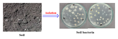
On arrival at the laboratory, the samples were freed of stones and plant fragments. A precisely weighed 1 g aliquot was placed in a sterile tube and combined with 9 mL of sterile distilled water. To dislodge microorganisms from the soil matrix, the suspension was agitated vigorously on a Vortex MX-S mixer for 5 min and then left undisturbed for 10 min so that coarser particles could settle. This preparation constituted the 10-¹ dilution. A 1 mL portion of the overlying liquid was next passed into a fresh tube holding 9 mL of sterile distilled water, and tenfold serial dilutions were continued as far as 10-5 [5]. From each dilution step, 100 µL of the bacterial suspension was distributed over the surface of sterile Petri dishes charged with a range of media by the spread-plate method. The media comprised Luria Bertani (LB) agar (yeast extract, 5 g/L; NaCl, 10 g/L; peptone, 10 g/L; agar, 15 g/L), Nutrient Agar (NA) (peptone, 5 g/L; HM Peptone B, 1.5 g/L; yeast extract, g/L; NaCl, 5 g/L; agar, 15 g/L), and Tryptic Soy Agar (TSA) (tryptone, 15 g/L; soy peptone, 5 g/L; NaCl, 5 g/L; agar, 15 g/L). Seeded plates were held at 28 °C for 24 – 48 h to permit colony formation [6]. The colonies that emerged varied markedly in appearance differing in size, outline, colour, margin, elevation, and surface. Discrete colonies were picked with care and streaked repeatedly onto fresh medium to yield pure cultures. Through successive subculturing, morphologically distinguishable colonies were separated and purified [7]. Each purified colony was taken to represent a single bacterial isolate and was retained for the ensuing morphological, biochemical, and molecular work [8, 9].
Identification of Bacterial Isolates by MALDI-TOF MS
For identification, pure cultures were grown for 24 h under suitable incubation conditions and then typed by matrix-assisted laser desorption/ionization time-of-flight mass spectrometry (MALDI-TOF MS). The resulting mass-spectral fingerprints were matched against the reference entries in the MALDI-TOF MS library to establish species identity [10]. Fresh colony material was applied directly onto a polished-steel MALDI target and overlaid with α-cyano-4-hydroxycinnamic acid (CHCA) matrix solution. Spectra were captured in linear positive-ion mode across a 2–20 kDa window, compared with the reference database in the MALDI Biotyper software, and interpreted per the manufacturer's score-based guidelines.
Evaluation of Antifungal Activity
Antifungal potential was first screened by the dual-culture antagonism assay. Five fungal test organisms were employed four Fusarium spp. strains and a single Alternaria sp. strain. The fungal pathogens were maintained on Potato Dextrose Agar (PDA) (potato extract, 200 g/L; glucose, 20 g/L; agar, 15 g/L), while the bacteria were propagated on NA. For the assay, bacterial isolates were laid down on the agar by the lawn-culture technique and then co-cultured with the fungal pathogens so that their inhibitory capacity could be assessed [11, 12]. Once the fungal colonies were fully grown, plugs bearing actively expanding mycelium were cut out aseptically with a sterile cork borer and moved onto fresh PDA plates. A 5 mm mycelial disc excised from the edge of a 7 day old culture was positioned some 25 – 30 mm from the plate centre under sterile conditions, and the bacterial strain was streaked on the far side, squarely opposite the fungal plug. All plates were incubated at 28 ± 2 °C for 7 days, alongside control plates carrying the fungal pathogen alone under matching conditions. After incubation, the zone of inhibition arising between the bacterial colony and the advancing mycelium was measured in millimetres to quantify antifungal activity [13]. To place the antagonism on a numerical footing, the radial growth inhibition (RI, %) of each pathogen was computed. This index expresses the shrinkage in fungal colony growth relative to the untreated control and is a widely adopted measure of microbial antagonistic efficacy. It was obtained from the following equation:
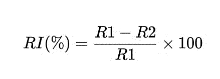
where R1 is the radius of the fungal colony on the control plate (mm), and R2 is the radius of the fungal colony measured toward the antagonistic bacterial colony on the treatment plate (mm). Every experiment was run in triplicate, and outcomes are reported as the mean ± standard deviation (SD).
Optimization of bioactive secondary metabolite production
To refine the yield of bioactive secondary metabolites from the most active strain, Paenibacillus lautus AD 83, the roles of medium composition, pH, and temperature were examined. The strain was propagated in five liquid media: Nutrient Broth (NB) (peptone, 5 g/L; yeast extract, 3 g/L; NaCl, 5 g/L), Luria Bertani (LB) broth, Tryptic Soy Broth (TSB), a nitrogen-free (N-free) broth (K2HPO4, 0.1 g/L; KH2PO4, 0.4 g/L; MgSO4, 0.2 g/L; NaCl, 0.1 g/L; CaCl2, 0.02 g/L), and an oat–corn broth (oat flour, 15 g/L; corn flour, 15 g/L; yeast extract, 3 g/L; NaCl, 5 g/L; CaCO3, 1 g/L). The impact of starting pH (5.0–8.0) and incubation temperature (20–40 °C) on metabolite output was likewise evaluated. The organism was grown under each condition and metabolite production quantified in mg/L. Medium pH was set prior to sterilization with 1 N HCl or 1 N NaOH. Temperature trials involved a 3-day incubation at each target temperature. All runs were carried out in triplicate. Following incubation, cultures were centrifuged and the metabolite concentration in the cell-free supernatant determined. From these data, the optimal medium, pH, and temperature for secondary-metabolite production by Paenibacillus lautus AD 83 were established.
Results and Discussion
Physicochemical analysis of the soil sample
Findings from the physicochemical assay of the soil are laid out in Table 1. Examination of the material taken from the 0–10 cm horizon showed the soil to be moderately saline. In the soil solution, sulfate (SO4²-) prevailed among the anions at 4.50 meq/L, trailed by chloride (Cl-) at 2.00 meq/L and bicarbonate (HCO3-) at 0.30 meq/L; the comparatively high sulfate and chloride figures point to a sulfatechloride type of salinity. On the cation side, sodium (Na+) dominated at 4.30 meq/L, with calcium (Ca²+) and magnesium (Mg²+) at 1.50 and 1.00 meq/L, respectively. This sodium predominance signals an accumulation of readily soluble salts and an advancing salinization process, which may impair soil structure by breaking down aggregates and lowering water infiltration and permeability.
The dry residue of the soil solution stood at 0.75 %, and the total soluble-salt content at 0.68 %, together confirming placement in the moderately saline class. A pH of 8.2 lent the soil a slightly alkaline character, potentially curbing the availability of certain nutrients phosphorus and micronutrients in particular to plants, while the electrical conductivity (EC) of 5.2 mS/cm reflected a fairly high load of soluble salts. Taken together, these attributes portray a moderately saline, mildly alkaline agroecological setting conditions apt to harbour stress-tolerant microorganisms able to make biologically active metabolites, and hence a promising well from which to draw microbes of agricultural and biotechnological interest.
Table 1.
Chemical characteristics of a moderately saline soil sample (0 – 10 cm depth)
|
Depth (cm) |
HCO₃⁻ (meq/100 g) |
Cl⁻ (meq/100 g) |
SO₄²⁻ (meq/100 g) |
Ca²⁺ (meq/100 g) |
Mg²⁺ (meq/100 g) |
Na⁺ (meq/100 g) |
Dry Residue (%) |
Total Soluble Salts (%) |
pH |
EC (mS/cm) |
|---|---|---|---|---|---|---|---|---|---|---|
| 0-10 | 0.30 | 2.00 | 4.50 | 1.50 | 1.00 | 4.30 | 0.75 | 0.68 | 8.2 | 5.2 |
Gram staining, microscopic observation, and morphological identification of bacterial isolates
Over the course of the study, more than fifteen bacterial isolates were retrieved from the collected soil. They were first told apart by colony traits—shape, margin, colour, surface texture, and size. Although the overall bacterial count in the sampled soil was rather modest, the bulk of the isolates presented as salt-tolerant organisms, in keeping with the moderately saline nature of the site. Preliminary morphological work, combined with Gram staining and microscopy, indicated that most isolates fell within the genus Bacillus: they proved to be Gram-positive rods, matching the hallmark features of Bacillus species. On the strength of the initial screening and morphological typing, six representative strains with distinctive colony characteristics were singled out for further identification and biological evaluation (Figure 1).
Identification of Bacterial Isolates by MALDI-TOF MS
Table 2.
Identification by MALDI-TOF MS
| Isolate code | Identified species | Identification score |
|---|---|---|
| AD80 | Bacillus atrophaeus | 2.31 |
| AD81 | Priestia endophytica | 2.27 |
| AD83 | Paenibacillus lautus | 2.34 |
| AD87 | Paenibacillus sp. | 2.18 |
| AD89 | Peribacillus frigoritolerans | 2.26 |
| AD90 | Bacillus atrophaeus | 2.29 |
As set out in Table 2, the MALDI-TOF MS analysis placed all six isolates chiefly within the family Bacillaceae. In detail, isolate AD80 was identified as Bacillus atrophaeus, AD81 as Priestia endophytica, AD83 as Paenibacillus lautus, AD87 as Paenibacillus sp., AD89 as Peribacillus frigoritolerans, and AD90 as Bacillus atrophaeus. The identification scores spanned 2.18 to 2.34, denoting dependable assignment of the isolates at the genus level. The organisms so identified are familiar constituents of the soil microbiota, many of them recognized for their capacity to spur plant growth and to yield biologically active metabolites with antifungal and antibacterial effects.
Evaluation of the antifungal activity of bacteria
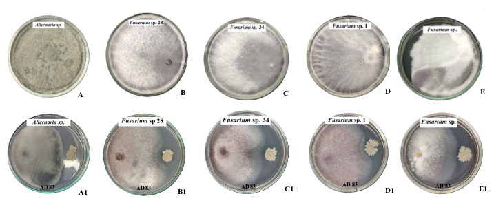
A-E - Control plates (fungi cultured alone): A - Alternaria sp.; B - Fusarium sp. 28; C - Fusarium sp. 34; D - Fusarium sp. 1; E - Fusarium sp.
A1-E1 - Dual-culture plates with Paenibacillus lautus AD 83: A1 - Alternaria sp. + P. lautus AD 83; B1 - Fusarium sp. 28 + P. lautus AD 83; C1 - Fusarium sp. 34 + P. lautus AD 83; D1 - Fusarium sp. 1 + P. lautus AD 83; E1 - Fusarium sp. + P. lautus AD 83.
Table 3.
Values
| № |
Phytopathogenic fungi |
Strain name |
Radial inhibition (RI %) |
| 1 |
Alternaria sp. |
Paenibacillus lautus AD 83 |
45 % |
| 2 |
Fusarium sp. 28 |
Paenibacillus lautus AD 83 |
34 % |
| 3 |
Fusarium sp. 34 |
Paenibacillus lautus AD 83 |
40 % |
| 4 |
Fusarium sp. 1 |
Paenibacillus lautus AD 83 |
38 % |
| 5 |
Fusarium sp. |
Paenibacillus lautus AD 83 |
36 % |
As the table shows, the top-performing strain, Paenibacillus lautus AD 83, held back every phytopathogenic fungus put to the test. The strongest suppression (45 %) fell on Alternaria sp., whereas the weakest (34 %) was registered against Fusarium sp. 28; inhibition of the remaining Fusarium isolates lay between 34 % and 40 %. These outcomes attest that P. lautus AD 83 wields pronounced antagonistic activity toward phytopathogenic fungi.
Effect of culture medium composition, ph, and temperature on secondary metabolite production
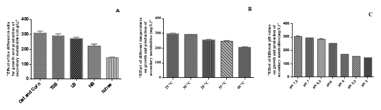
How pH, temperature, and the choice of carbon and nitrogen sources bear on the output of biologically active secondary metabolites by soil bacteria has been the subject of extensive study [14]. Bamola et al. [15] for instance, worked out the growth conditions best suited to members of the genus Bacillus for maximizing growth as well as antidermatophytic and antifungal activity; they found the optimum temperature for metabolite formation to be 37 °C and the optimal pH to fall between 7.0 and 8.5, under which an inhibition zone as large as 28 mm was recorded. Comparable observations were reported by Rustamova et al. [16]. In the present investigation, the best culturing conditions for Paenibacillus lautus AD 83 were pinned down. The strain turned out 296 mg/L of secondary metabolites at 25 °C. Among the media assessed, oat corn broth and TSB backed the highest yields, delivering 312 mg/L and 295 mg/L, respectively. The optimal pH for metabolite formation proved to be 7.5, at which the strain produced 303 mg/L.
Conclusion
In summary, more than fifteen bacterial isolates were recovered from moderately saline, slightly alkaline agroecological soil near Khonobod village, Kashkadarya Region. MALDI-TOF MS placed six representative strains within the family Bacillaceae, spanning Bacillus atrophaeus, Priestia endophytica, Paenibacillus lautus, Paenibacillus sp., and Peribacillus frigoritolerans, with identification scores between 2.18 and 2.34. Screening by the dual-culture assay singled out Paenibacillus lautus AD 83 as the most effective antagonist, curbing the radial growth of the tested Alternaria and Fusarium pathogens by 34–45 %. For this strain, the conditions most conducive to secondary-metabolite production were an oat–corn medium, 25 °C, and pH 7.5, yielding up to 312 mg L-¹. Collectively, these results identify Paenibacillus lautus AD 83 as a promising microorganism for the biological control of phytopathogenic fungi and for the development of environmentally friendly biofungicides.
References