Organ-specific distribution of flavonoids and free amino acids in Astragalus campylorrhynchus
УДК 581.19+543.544
Abstract
This study comparatively investigated the organ-specific distribution of flavonoids and free amino acids in different morphological parts of Astragalus campylorrhynchus Fisch. & C.A.Mey. using high-performance liquid chromatography. Roots, stems and leaves, seeds, and seed coats were analyzed. The qualitative and quantitative composition of flavonoids was determined by HPLC-DAD, revealing distinct differences in the distribution of individual compounds among the investigated organs. Rutin was detected in all analyzed samples, with the highest concentrations found in stems and leaves (113.231 mg/100 g) and seeds (110.846 mg/100 g), followed by roots (90.231 mg/100 g) and seed coats (41.716 mg/100 g). The seed coat was characterized by relatively high levels of apigenin and quercetin. Free amino acids were determined by HPLC-DAD following pre-column derivatization with phenyl isothiocyanate. A total of 20 free amino acids were quantified. Their total content was 96.167 mg/g in stems and leaves, 60.073 mg/g in seed coats, 49.565 mg/g in seeds, and 29.524 mg/g in roots. Proline was particularly abundant in stems and leaves (21.604 mg/g), whereas serine predominated in the seed coat (26.650 mg/g). The results demonstrate that the vegetative and generative organs of A.campylorrhynchus exhibit distinct chemical profiles with respect to the qualitative and quantitative composition of flavonoids and free amino acids. These findings expand the phytochemical characterization of A.campylorrhynchus and provide a basis for further targeted studies of its individual morphological parts.
Аннотация
В настоящем исследовании методом высокоэффективной жидкостной хроматографии проведено сравнительное изучение органоспецифического распределения флавоноидов и свободных аминокислот в различных морфологических частях Astragalus campylorrhynchus Fisch. & C.A.Mey. В качестве объектов исследования использовали корни, стебли и листья, семена и семенную оболочку. Качественный и количественный состав флавоноидов определяли методом ВЭЖХ-ДМД. Установлено неравномерное распределение индивидуальных флавоноидов между исследованными органами. Рутин был обнаружен во всех анализируемых образцах; его максимальное содержание установлено в стеблях и листьях (113,231 мг/100 г) и семенах (110,846 мг/100 г), тогда как в корнях и семенной оболочке его содержание составляло 90,231 и 41,716 мг/100 г соответственно. Семенная оболочка характеризовалась относительно высоким содержанием апигенина и кверцетина. Свободные аминокислоты определяли методом ВЭЖХ-ДМД после предколоночной дериватизации фенилизотиоцианатом. Количественно определено 20 свободных аминокислот. Их суммарное содержание составило 96,167 мг/г в стеблях и листьях, 60,073 мг/г в семенной оболочке, 49,565 мг/г в семенах и 29,524 мг/г в корнях. В стеблях и листьях отмечено высокое содержание пролина (21,604 мг/г), тогда как семенная оболочка характеризовалась преобладанием серина (26,650 мг/г). Полученные результаты свидетельствуют о дифференциации химического профиля вегетативных и генеративных органов A.campylorrhynchus по качественному и количественному составу флавоноидов и свободных аминокислот. Полученные данные расширяют фитохимическую характеристику вида и могут служить основой для дальнейших целенаправленных исследований его отдельных морфологических частей.
Keywords: Astragalus campylorrhynchus; flavonoids; free amino acids; organ-specific distribution; HPLC-DAD; rutin; proline.
Ключевые слова: Astragalus campylorrhynchus; флавоноиды; свободные аминокислоты; органоспецифическое распределение; ВЭЖХ-ДМД; рутин; пролин.
Introduction
The genus Astragalus L. (Fabaceae) is one of the largest plant genera and is characterized by considerable phytochemical diversity. Its representatives contain numerous primary and secondary metabolites, including flavonoids and isoflavonoids, triterpenoid saponins, polysaccharides, and other phenolic compounds [2; 6; 17]. Although the phytochemistry of medicinal species, particularly A.membranaceus, has been extensively investigated [6; 17], the organ-specific distribution of metabolites remains insufficiently characterized in many Astragalus species.
Flavonoids are among the important secondary metabolites of Astragalus and include flavones, flavonols, isoflavones, isoflavans, pterocarpans, and their glycosides [2]. Their qualitative and quantitative profiles may vary among plant organs due to differences in physiological function, developmental stage, and environmental conditions. Flavonoids have been reported in the stems and leaves of A.membranaceus [4], while chromatographic studies of A.mongholicus and A.membranaceus roots have identified calycosin, formononetin, ononin, and other isoflavonoids [14]. These findings support the separate evaluation of morphological parts when characterizing Astragalus phytochemistry. High-performance liquid chromatography coupled with diode-array detection (HPLC-DAD) is particularly suitable for this purpose because it combines chromatographic separation with retention and UV spectral characteristics [14].
Organ-specific chemical differentiation also involves primary metabolites. Free amino acids participate in protein biosynthesis, carbon and nitrogen metabolism, plant growth, development, and other cellular processes [8]. Their distribution may vary according to the metabolic functions of individual tissues. The close integration of photosynthesis, organic acid metabolism, the tricarboxylic acid cycle, and amino acid biosynthesis further contributes to metabolic differentiation among plant organs [13]. Therefore, comparative analysis of free amino acids can complement flavonoid profiling in evaluating organ-specific chemical characteristics. For their chromatographic determination, pre-column derivatization with phenyl isothiocyanate (PITC), followed by reversed-phase HPLC, provides effective separation and quantitative analysis of individual amino acids [3].
Parallel investigation of flavonoids and free amino acids enables organ-specific chemical differentiation to be assessed at the levels of secondary and primary metabolism, respectively [6; 8; 13]. However, their simultaneous accumulation within an organ should not be interpreted as evidence of a direct correlation or causal relationship without appropriate biological replication and statistical or metabolomic analyses. In the present study, these metabolite groups were therefore considered complementary indicators of organ-specific chemical profiles.
Astragalus campylorrhynchus Fisch. & C.A.Mey. occurs in the flora of Uzbekistan, particularly in loess and rocky foothill habitats, and exhibits ephemeral-xerophytic ecological characteristics. Flowering generally occurs in April–May, followed by fruiting in May-June [16]. Compared with extensively studied species such as A.membranaceus and A.mongholicus, however, its phytochemical composition remains poorly characterized. In particular, comparative quantitative information on flavonoids and free amino acids in its roots, stems and leaves, seeds, and seed coats is limited.
Therefore, the aim of this study was to determine the qualitative and quantitative composition of individual flavonoids and free amino acids in the roots, stems and leaves, seeds, and seed coats of A.campylorrhynchus using high-performance liquid chromatography and to comparatively evaluate their organ-specific distribution based on primary and secondary metabolite profiles.
Materials and methods
Plant material and sample preparation. Roots, stems and leaves, seeds, and seed coats of A.campylorrhynchus Fisch. & C.A.Mey. (Fabaceae) were collected on 24 April 2025 near the Orzu railway station, Kurama Range, Namangan Region, Uzbekistan. Botanical identification was based on morphological and taxonomic characteristics. The collections were registered under Nos. 56–58, and a voucher specimen was deposited in the herbarium of the Department of Biology, Kokand State University (KD000247).
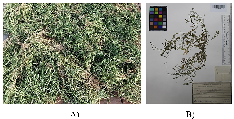
Plant materials were cleaned, separated into morphological parts, and dried under laboratory conditions. Samples for flavonoid analysis were ground to 0.1-1.5 mm. The collected plant material and its voucher specimen are shown in Figure 1.
Extraction of flavonoids. Each dried and ground sample (1.0000 g) was extracted with 99 mL of 70 % ethanol under reflux at 50–60 °C for 2 h with continuous stirring. The mixture was then sonicated at 35 °C for 15 min, followed by a 10-min interval and a second 15-min sonication. After cooling, the extract was filtered; 2 mL of filtrate was passed through a 0.2-µm membrane filter. A 100-µL aliquot was diluted with chromatographic eluent to 1.0 mL for HPLC-DAD analysis.
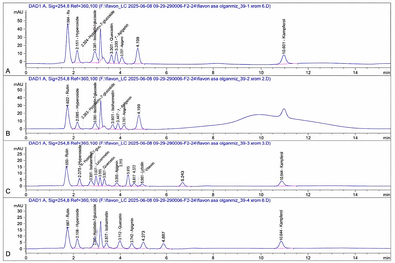
HPLC-DAD analysis of flavonoids. Flavonoids were analyzed using an Agilent Technologies 1260 HPLC-DAD system with an Eclipse XDB-C18 column (250 × 4.6 mm, 5 µm). The mobile phase was acetonitrile–buffer (pH 2.92), 30:70 (v/v), under isocratic conditions at 0.75 mL/min. The analysis time was 20 min and the injection volume 5 µL. DAD spectra were recorded at 200–400 nm, with detection at 254, 320, and 381 nm. Compounds were identified by comparison of retention times and DAD spectra with reference standards and quantified as mg/100 g dry weight.
Extraction and PITC derivatization of free amino acids. Proteins and peptides were precipitated by mixing 1 mL of aqueous plant extract with 1 mL of 20 % trichloroacetic acid (TCA). After 10 min, the mixture was centrifuged at 8000 rpm for 15 min, and 0.1 mL of supernatant was lyophilized. The residue was dissolved in triethylamine–acetonitrile–water (1:7:1, v/v/v) and dried twice to neutralize residual acid. Pre-column derivatization with phenyl isothiocyanate (PITC) was performed according to Cohen and Strydom [3].
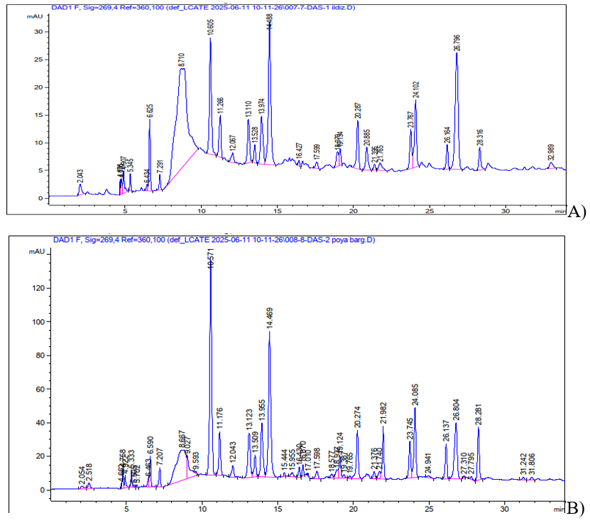
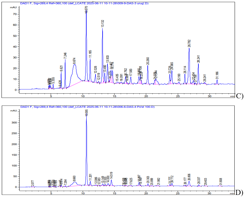
HPLC-DAD analysis of free amino acids. Free amino acids were analyzed using an Agilent Technologies 1200 HPLC-DAD system with a Discovery HS-C18 column (75 × 4.6 mm). Mobile phase A was 0.14 M CH3COONa containing 0.05 % triethylamine (pH 6.4), and mobile phase B was acetonitrile. The flow rate was 1.2 mL/min and detection was performed at 269 nm. The gradient was: 0–2.5 min, 1–6 % B; 2.51–40.0 min, 6–30 % B; 40.1–45.0 min, 30–60 % B; 45.1–50.0 min, 60 % B; and 50.1–55.0 min, 60–0 % B. Amino acids were identified by comparison with the reference amino acid mixture and quantified as mg/g dry weight.
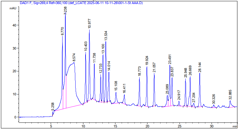
The reference chromatogram was used to identify individual amino acids based on their retention times.
Calculation and comparative evaluation.Organ-specific distributions were evaluated by comparing individual metabolite concentrations and their summed values. The sum of quantified flavonoids was calculated as ΣF = ΣCi, where Ci is the concentration of each individually quantified flavonoid (mg/100 g dry weight). ΣF was not interpreted as total flavonoid content (TFC), because it included only compounds quantified using available reference standards.
Similarly, total quantified free amino acids were calculated as ΣFAA = ΣCi and expressed as mg/g dry weight. Because dispersion data from biological replicates were unavailable, differences among organs were evaluated descriptively and were not interpreted in terms of statistical significance.
Results and discussion
Organ-specific distribution of flavonoids in A.campylorrhynchus. HPLC-DAD revealed clear qualitative and quantitative differences among the investigated organs. Six flavonoids—apigenin, rutin, hypolaetin-7-O-D-glucoside, hyperoside, isorhamnetin, and quercetin—were quantified. Rutin occurred in all organs and was highest in stems and leaves (113.231 mg/100 g) and seeds (110.846 mg/100g), followed by roots (90.231 mg/100 g) and seed coats (41.716 mg/100 g) (Table 1).
Table 1.
Quantitative composition of individual flavonoids in different organs of A.campylorrhynchus, mg/100 g dry weight
| № | Flavonoid | Root | Stems and leaves | Seed | Seed coat |
|---|---|---|---|---|---|
| 1 | Apigenin | – | 0.617 | – | 4.561 |
| 2 | Rutin | 90.231 | 113.231 | 110.846 | 41.716 |
| 3 | Hypolaetin 3-7-O-D-glucoside | – | – | 0.111 | 0.423 |
| 4 | Hyperoside | – | – | 0.045 | – |
| 5 | Isorhamnetin | – | 0.841 | – | 0.324 |
| 6 | Quercetin | 14.348 | 14.865 | – | 15.863 |
| ΣF | 104.579 | 129.554 | 111.002 | 62.887 |
Note: “–” indicates that the compound was not quantitatively detected in the corresponding sample; ΣF represents the sum of individual flavonoids quantified using the available reference standards.
The highest ΣF occurred in stems and leaves (129.554 mg/100 g), followed by seeds (111.002 mg/100 g), roots (104.579 mg/100 g), and seed coats (62.887 mg/100 g). Rutin represented approximately 87.4 % of ΣF in stems and leaves and more than 99.9 % in seeds.
This organ-dependent distribution agrees with previous studies of Astragalus. Differences among plant parts have been demonstrated by UPLC-MS/MS [10], while HPLC-DAD-ELSD studies showed distinct flavonoid profiles between stems and leaves and roots of Huang-qi [11]. Development-dependent flavonoid accumulation and differential expression of flavonoid biosynthetic genes have also recently been demonstrated in the stems and leaves of A. membranaceus [5], from whose stems and leaves 11 flavonoids, including three new glycosides, were isolated [15].
The profile of A.campylorrhynchus also differed from those reported for other Astragalus species. Calycosin, formononetin, ononin, and related isoflavonoids have been described in A.membranaceus and A.mongholicus roots [14; 12], whereas rutin predominated in the present samples. Rutin has also been reported as a major polyphenol in organs of A.campylosema, while hyperoside predominated in some organs of A.hirsutus [9], supporting both species- and organ-dependent variation.
Quercetin concentrations were similar in roots, stems and leaves, and seed coats (14.348–15.863 mg/100 g), whereas apigenin was approximately 7.4-fold higher in seed coats (4.561 mg/100 g) than in stems and leaves (0.617 mg/100 g). Thus, the organ with the highest ΣF did not necessarily contain the highest concentration of every individual flavonoid. Because extraction and analytical conditions vary among studies, comparison of dominant compounds and distribution patterns is more appropriate than direct comparison of absolute concentrations. Organ-specific distribution of free amino acids in A.campylorrhynchus.
Twenty free amino acids were quantitatively determined. ΣFAA was highest in stems and leaves (96.167 mg/g), followed by seed coats (60.073 mg/g), seeds (49.565 mg/g), and roots (29.524 mg/g) (Table 2).
Table 2.
Quantitative composition of free amino acids in different organs of A.campylorrhynchus, mg/g dry weight
| № | Amino acid | Root | Stems and leaves | Seed | Seed coat |
|---|---|---|---|---|---|
| 1 | Aspartic acid | 2.950 | 4.257 | 2.912 | 2.146 |
| 2 | Glutamic acid | 0.420 | 1.792 | 3.800 | 0.773 |
| 3 | Serine | 1.919 | 11.235 | 5.728 | 26.650 |
| 4 | Glycine | 0.574 | 1.789 | 1.550 | 1.278 |
| 5 | Asparagine | 0.549 | 1.804 | 1.559 | 1.271 |
| 6 | Glutamine | 0.366 | 1.164 | 7.242 | 0.822 |
| 7 | Cysteine | 0.156 | 0.682 | 1.346 | 0.245 |
| 8 | Threonine | 1.151 | 4.480 | 1.045 | 1.523 |
| 9 | Arginine | 0.131 | 0.608 | 0.929 | 0.286 |
| 10 | Alanine | 1.665 | 5.407 | 2.361 | 2.019 |
| 11 | Proline | 6.922 | 21.604 | 2.300 | 7.633 |
| 12 | Tyrosine | 0.270 | 1.353 | 0.833 | 2.365 |
| 13 | Valine | 1.229 | 4.372 | 2.404 | 1.484 |
| 14 | Methionine | 0.454 | 5.462 | 2.216 | 0.895 |
| 15 | Histidine | 3.258 | 11.377 | 2.158 | 2.228 |
| 16 | Isoleucine | 1.060 | 3.155 | 1.297 | 1.227 |
| 17 | Leucine | 2.352 | 7.109 | 2.397 | 2.316 |
| 18 | Tryptophan | 3.473 | 5.067 | 5.163 | 3.773 |
| 19 | Phenylalanine | 0.463 | 3.282 | 1.972 | 1.005 |
| 20 | Lysine | 0.162 | 0.171 | 0.352 | 0.133 |
| ΣFAA | 29.524 | 96.167 | 49.565 | 60.073 |
Stems and leaves were characterized by high proline (21.604 mg/g), histidine (11.377 mg/g), and serine (11.235 mg/g), with proline accounting for approximately 22.5 % of ΣFAA. Proline also predominated in roots (6.922 mg/g). Seeds contained relatively high glutamine (7.242 mg/g), serine (5.728 mg/g), and tryptophan (5.163 mg/g), whereas serine predominated in seed coats (26.650 mg/g), representing approximately 44 % of ΣFAA.
These differences may reflect the distinct metabolic functions of individual organs because amino acids participate in protein biosynthesis, carbon–nitrogen metabolism, growth, and development [8; 13]. The high proline level in stems and leaves is noteworthy because increased proline accumulation has been reported in A.tennesseensis under water stress [1]. However, since stress and control treatments were not compared here, the observed proline level cannot be interpreted as direct evidence of stress tolerance.
Comparative organ-specific profiling of flavonoids and free amino acids
Combined evaluation demonstrated distinct chemical profiles among the organs. Stems and leaves had the highest ΣF (129.554 mg/100 g) and ΣFAA (96.167 mg/g). Seeds showed a high ΣF (111.002 mg/100 g), dominated by rutin, whereas roots had 104.579 mg/100 g ΣF but the lowest ΣFAA (29.524 mg/g). Seed coats were distinguished by comparatively high apigenin, quercetin, and serine levels.
These differences are consistent with organ-dependent metabolite profiles reported in other Astragalus species [5; 7; 11; 15]. Nevertheless, rutin predominance in the stems and leaves and seeds of A.campylorrhynchus should be regarded as a characteristic of the investigated species and samples rather than a general feature of the genus, which exhibits substantial phytochemical diversity [10].
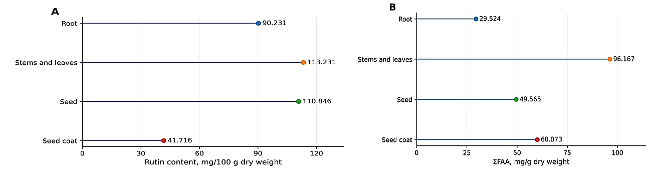
The simultaneous high ΣF and ΣFAA values in stems and leaves do not demonstrate a direct relationship between the two metabolite groups; establishing such an association would require biological replicates, correlation analysis, and broader metabolomic investigation. Accordingly, flavonoids and free amino acids were considered complementary indicators of the organ-specific chemical profile of A.campylorrhynchus.
Overall, stems and leaves were distinguished by the highest ΣF and ΣFAA values, seeds by pronounced rutin predominance, and seed coats by comparatively high apigenin, quercetin, and serine levels. These findings demonstrate the importance of treating individual morphological parts of A.campylorrhynchus as separate analytical objects. Since biological activity was not evaluated, the observed profiles should not be directly interpreted in terms of antioxidant, pharmacological, or nutritional efficacy.
Conclusion
The organ-specific distribution of flavonoids and free amino acids in the roots, stems and leaves, seeds, and seed coats of Astragalus campylorrhynchus Fisch. & C.A.Mey. was comparatively characterized using HPLC-DAD. The results demonstrated distinct qualitative and quantitative metabolite profiles among the vegetative and generative organs.
Rutin was the predominant quantified flavonoid, reaching its highest levels in stems and leaves (113.231 mg/100 g) and seeds (110.846 mg/100 g). The sum of individually quantified flavonoids (ΣF) was highest in stems and leaves (129.554 mg/100 g), while seed coats were distinguished by comparatively high levels of apigenin and quercetin.
A total of 20 free amino acids were quantified. The ΣFAA values were 96.167 mg/g in stems and leaves, 60.073 mg/g in seed coats, 49.565 mg/g in seeds, and 29.524 mg/g in roots. Proline was particularly abundant in stems and leaves, whereas serine predominated in seed coats.
These findings demonstrate the organ-specific differentiation of primary and secondary metabolite profiles in A.campylorrhynchus and expand the available phytochemical information on this species. The results also support the consideration of individual morphological parts as separate analytical objects in further phytochemical studies and provide a basis for subsequent targeted chemical, metabolomic, and biological investigations.
References