Immobilization of alizarin red s onto silk fibroin fibers and spectral characterization
УДК 543.42:544.77:677.074
Abstract
In the present study, Alizarin Red S (ARS) was immobilized onto silk fibroin fibers with the aim of developing a novel functional composite material based on a natural biopolymer matrix. The physicochemical properties of the resulting ARS-silk fibroin system were investigated using diffuse reflectance spectroscopy (DRS) and Fourier transform infrared (FTIR) spectroscopy. The results demonstrated that the optimal immobilization conditions were achieved within the pH range of 3.32 – 4.65 and at temperatures between 25 and 30 °C. Under these conditions, 0.2 g of silk fibroin effectively immobilized up to 79.9 mmol·g-¹ of ARS within 30 min. The appearance of a characteristic absorption band at 435 – 440 nm in the DRS spectra confirmed the successful immobilization of ARS onto the fibroin matrix. FTIR analysis further indicated the presence of intermolecular interactions between the amide groups of silk fibroin and the hydroxyl and sulfonate groups of ARS molecules. The obtained findings demonstrate that the ARS-modified silk fibroin possesses excellent sorption properties and can be considered a promising functional biomaterial for the development of optical and colorimetric sensors intended for the determination of heavy metal ions in environmental and analytical applications.
Аннотация
В данном исследовании с целью создания нового функционального композитного материала на основе природного биополимера – волокон шелкового фиброина – была проведена иммобилизация реагента Ализарин красный S (АКС). Физико-химические свойства полученной системы Ализарин красный S (АКС)-фиброин были исследованы методами спектроскопии диффузного отражения (DRS) и инфракрасной спектроскопии с преобразованием Фурье (FTIR). Результаты исследования показали, что оптимальные условия процесса иммобилизации достигаются в диапазоне pH 3,32 – 4,65 при температуре 25–30 °C. При указанных условиях образец шелкового фиброина массой 0,2 г способен эффективно связывать до 79,9 ммоль·г-¹ молекул АКС в течение 30 минут. Характерная полоса поглощения в области 435 – 440 нм, наблюдаемая в спектрах DRS, подтверждает успешную иммобилизацию АКС на матрице фиброина. Анализ FTIR-спектров свидетельствует о наличии межмолекулярных взаимодействий между амидными группами фиброина и гидроксильными и сульфонатными группами АКС. Полученные результаты демонстрируют, что модифицированный Ализарин красный S (АКС) шелковый фиброин обладает высокими сорбционными свойствами и может рассматриваться как перспективный функциональный биоматериал для создания оптических и колориметрических сенсоров, предназначенных для определения ионов тяжелых металлов.
Keywords: Alizarin Red S; silk fibroin; immobilization; diffuse reflectance spectroscopy (DRS); biomaterial; optical sensor.
Ключевые слова: ализариновый красный S; фиброин шелка; иммобилизация; спектроскопия диффузного отражения (DRS); биоматериал; оптический датчик.
1. Introduction
In recent years, the immobilization of organic indicator reagents onto solid support materials has become one of the rapidly developing scientific directions in analytical chemistry, biomaterials, and optical sensor technologies [1–3]. Immobilization technology makes it possible to enhance the chemical and photochemical stability of indicator reagents, reduce their leaching from the support matrix, improve reusability, and significantly enhance analytical parameters such as sensitivity and selectivity [1]. For this reason, extensive research is being conducted on the immobilization of various chromogenic and complex-forming reagents onto support materials based on silica gel, chitosan, cellulose, polymer membranes, nanocomposites, and natural biopolymers [2,3]. Modern research demonstrates that optical sensors based on immobilized reagents exhibit high analytical performance in environmental monitoring, biological fluid analysis, food safety, and industrial process control. Therefore, combining immobilization technologies with novel functional biomaterials is currently regarded as one of the priority scientific directions in analytical chemistry and sensor technologies.
Silk fibroin is a biodegradable material that induces a low-level inflammatory response in living organisms [4]. Due to its high biocompatibility, mechanical strength, controllable biodegradation properties, and excellent optical characteristics, it is widely applied as a promising biomaterial in various biomedical fields [5,6]. SF degrades at a controllable rate under in vivo and in vitro conditions [6]. The final degradation products consist of amino acids, which are easily assimilated by the organism [7]. These very properties are among the main reasons for the widespread application of silk fibroin in biomedicine [4]. Furthermore, by altering the degree of crystallinity and the β-sheet content of fibroin, as well as by processing it through various technological or post-treatment methods, its biodegradation rate can be controlled from several weeks to several years [8].
Silk fibroin obtained from local silk waste has been used as a functional support material, and the possibilities of immobilizing various organic analytical reagents, determining optimal conditions, and detecting Co(II) ions by sorption-spectroscopic methods have been studied. In particular, the successful immobilization of Eriochrome Black T reagent onto fibroin fiber, the immobilization mechanism, and the DRS and FTIR spectral characteristics of the resulting material were investigated, and this system was recommended as a promising material for the development of optical sensors [9].
However, an analysis of the available literature shows that the immobilization of ARS onto natural silk fibroin fiber, the optimal conditions of the immobilization process, and the diffuse reflectance and infrared spectral properties of the resulting material have not been sufficiently studied. This situation indicates the necessity of conducting additional research on the development of new functional biomaterials and optical sensors based on silk fibroin.
2. Experimental
2.1 Analytical Reagent Solutions
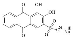
In this study, Alizarin Red S (ARS) was used as the analyte reagent to be immobilized. Alizarin Red S (CAS: 130-22-3) is an organic chromogenic reagent belonging to the class of anthraquinone derivatives, and its systematic name is sodium 3,4-dihydroxy-9,10-dioxo-9,10-dihydroanthracene-2-sulfonate. The reagent was purchased from Maclin (China). A standard solution of ARS at 1.0×10-³ mol/L was prepared, and from it, a working solution of 1.0×10⁻⁴ mol/L was obtained by dilution with distilled water. The prepared solutions were used for immobilization onto silk fibroin and for conducting spectrophotometric and diffuse reflectance spectroscopic (DRS) studies. The molecular structure of Alizarin Red S is shown in Figure 1.
2.2 Preparation of Acetate Buffer Solution
In the experiments, an acetate buffer solution was used to create a medium in the pH range of 3.0–6.0. The buffer solution was prepared by mixing 0.1 mol/L acetic acid (CH3COOH) and 0.1 mol/L sodium acetate (CH3COONa) solutions in appropriate proportions [10]. The required pH values were selected according to the Henderson–Hasselbalch equation and monitored using a pH meter. All solutions were prepared using analytically pure reagents and double-distilled water.
2.3 Preparation of the Polymer Matrix
Silk fibroin fiber isolated from local silk waste was used as the polymer matrix. The silk waste was initially cleaned of mechanical impurities and washed with distilled water. For the removal of the sericin layer (degumming), the sample was boiled in a 0.5 % (w/v) sodium carbonate (Na2CO3) solution for 30 min. Thereafter, the fibers were washed several times with distilled water until a neutral pH was achieved and dried at 40–50 °C until a constant mass was obtained. The prepared silk fibroin fiber was used as the polymer matrix in subsequent immobilization experiments [11,12].
2.4 Research Methods and Equipment
All experimental studies were carried out using modern analytical instrumentation. The mass of reagents and samples was measured on an AS 220.R2 Plus (Radwag, Poland) analytical balance with an accuracy of ±0.1 mg. Deionized water used in the experiments was obtained using an EASYpure RoDI (Thermo Scientific, USA) water purification system. The pH values of the solutions were determined using a FiveEasy F20 (Mettler Toledo, Switzerland) pH meter. The absorption spectra and optical density of Alizarin Red S solutions were studied using SPECORD 50 (Analytik Jena, Germany) and UV-1900i (Shimadzu, Japan) UV–Vis spectrophotometers. The diffuse reflectance spectra (DRS) of the immobilized Alizarin Red S–silk fibroin system were recorded on an Eye-One Pro (i1 Pro, X-Rite, Switzerland) mini-spectrophotometer in the wavelength range of 380–730 nm. FTIR spectra were obtained using an IRTracer-100 infrared Fourier-transform spectrometer (LabSolutions IR, Shimadzu Corporation, Japan) to identify the functional groups of silk fibroin and the immobilized material and the interactions between them. During immobilization experiments, a magnetic stirrer and a laboratory shaker were used for mixing solutions, and a thermostated laboratory heater was used for temperature control. The obtained spectral results were used to confirm the successful immobilization of Alizarin Red S onto the silk fibroin matrix and the interactions that occurred between functional groups during the immobilization process.
3. Results and Discussion
3.1 Efficiency of Alizarin Red S Immobilization onto the Fibroin Matrix
To evaluate the efficiency of the immobilization process, the degree of binding of Alizarin Red S (ARS) onto the surface of silk fibroin fiber was studied by the spectrophotometric method. The immobilization efficiency was evaluated by comparing the absorption spectra and optical density values of the reagent solution before and after immobilization.
3.1.1 Evaluation of Immobilization Efficiency
The immobilization efficiency (R, %) was calculated based on the optical density values of the Alizarin Red S solution before (A0) and after (A) immobilization using the following equation [13,14]:
where A0 is the optical density of the Alizarin Red S solution before immobilization, and A is the optical density of the Alizarin Red S solution after immobilization.
As a result of the calculation, the immobilization efficiency of Alizarin Red S onto silk fibroin fiber was found to be 73.6 %. This result indicates that the major portion of the reagent molecules was successfully transferred from the solution phase to the fibroin matrix.
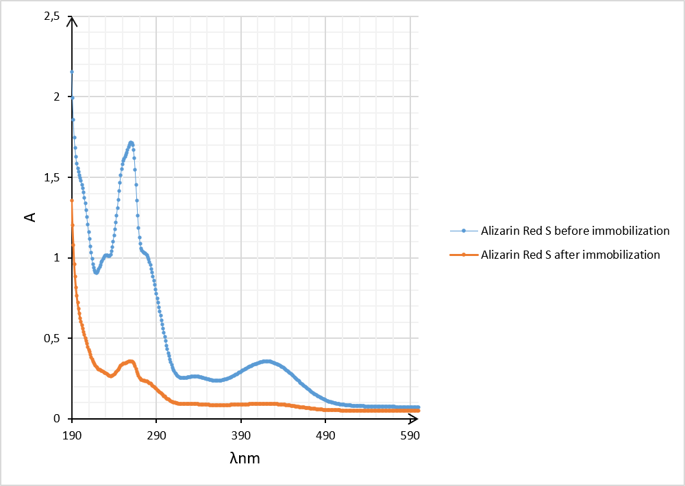
Comparison of the spectra shows that the concentration of the reagent in the solution phase decreased significantly during the immobilization process. The results of spectral analysis showed that before immobilization, the Alizarin Red S solution exhibited a maximum optical density of 0.356 at λmax = 420 nm, which decreased to 0.094 after immobilization. The approximately 73.6 % decrease in optical density confirms that the reagent molecules were efficiently adsorbed from the solution phase onto the surface of the fibroin fiber and immobilized.
The immobilization of Alizarin Red S onto silk fibroin fiber proceeded with high efficiency in the pH range of 2.15–6.65. This is explained by the influence of the pH of the medium on the degree of ionization of the functional groups of the reagent and fibroin, as well as their mutual interaction.
The pH of the medium is an important factor determining the immobilization efficiency of Alizarin Red S onto silk fibroin fiber. Changes in pH affect the degree of ionization of the functional groups of the reagent and fibroin, significantly influencing the course of the immobilization process. The results of the study are presented in Table 1.
Table 1.
Effect of pH on the immobilization of Alizarin Red S onto silk fibroin fiber [λmax = 430 nm; t = 20 ± 5 °C]
| Buffer solution | pH | R (%) |
|---|---|---|
| Acetate | 2.45 | 52.8 |
| 3.32 | 56.2 | |
| 4.65 | 56.5 | |
| 5.56 | 54.5 | |
| 6.65 | 60.0 | |
| Citrate | 3.32 | 62.5 |
| 4.65 | 73.6 | |
| 5.56 | 65.6 | |
| 6.65 | 66.3 | |
| 7.78 | 56.0 | |
| Ammonia | 8.45 | 32.0 |
| 9.18 | 36.0 | |
| 10.15 | 48.0 | |
| 11.09 | 49.0 | |
| 12.05 | 33.0 | |
| 12.80 | 31.0 |
As can be seen from Table 1, the immobilization efficiency of Alizarin Red S onto silk fibroin fiber depends significantly on the pH of the medium. The highest immobilization value was observed at pH 4.65, reaching 73.6 %. When the pH was increased beyond this value, the immobilization efficiency gradually decreased, falling in the range of 31–49 % in alkaline media. This is explained by changes in the degree of ionization of the functional groups in the fibroin and Alizarin Red S molecules, as well as the weakening of hydrogen bonds and electrostatic interactions between them. Therefore, pH 4.65 was selected as the optimal medium for all subsequent experiments.
After determining the optimal immobilization conditions, further studies were carried out on the silk fibroin fiber immobilized with Alizarin Red S. For this purpose, the diffuse reflectance (DRS) spectrum of the immobilized sample and the infrared Fourier-transform (FTIR) spectra were investigated to evaluate the binding of the reagent to the fibroin matrix and the spectral characteristics of the resulting material.
3.2 Spectral Characterization of Silk Fibroin Fiber Immobilized with Alizarin Red S
After determining the optimal immobilization conditions, the spectral characteristics of the silk fibroin fiber immobilized with Alizarin Red S were studied using diffuse reflectance spectroscopy (DRS) and UV–Vis spectrophotometry. Spectral analysis confirmed the successful implementation of the immobilization process and the binding of the reagent to the fibroin matrix.
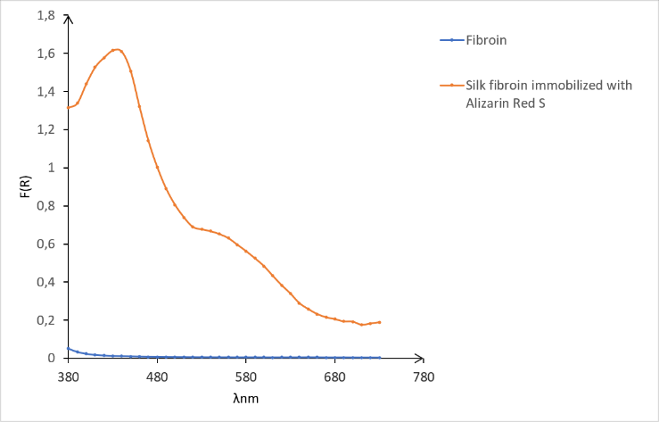
Figure 4 presents the diffuse reflectance (DRS) spectra of the initial silk fibroin fiber and the silk fibroin fiber immobilized with Alizarin Red S. The initial fibroin sample exhibited an almost constant and very low reflectance intensity over the entire investigated range of 380–730 nm, which is explained by the absence of characteristic chromophore groups in fibroin in the visible region [15,16]. However, after the immobilization of Alizarin Red S, the DRS spectrum changed drastically, and a distinct spectral maximum was observed in the 430–440 nm region. This maximum is associated with electronic transitions (π→π* and partially n→π* transitions) characteristic of the anthraquinone chromophore system of the Alizarin Red S molecule, confirming the successful immobilization of the reagent onto the fibroin surface. At the same time, the gradual decrease in reflectance intensity in the 500–700 nm range indicates an enhanced light interaction of the immobilized layer.
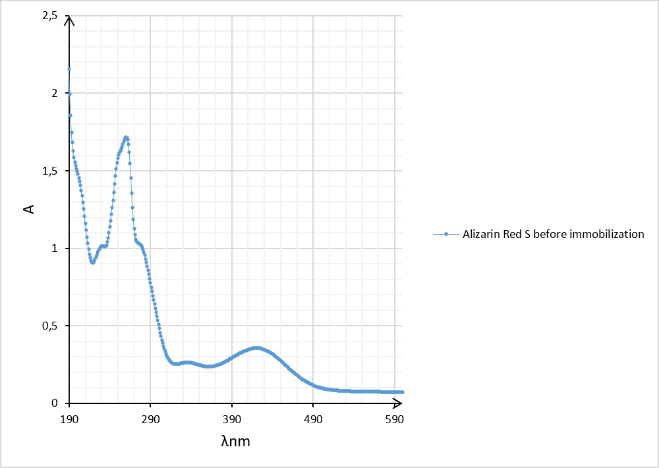
Figure 3 presents the UV–Vis absorption spectrum of the Alizarin Red S solution. The reagent exhibited a strong absorption maximum around 430 nm. This maximum coincides almost exactly with the maximum observed in the 430–440 nm range of the DRS spectrum. The correspondence of the spectral maxima indicates that the chromophore system of the Alizarin Red S molecule did not undergo significant changes during the immobilization process and that the optical activity of the reagent was preserved in the fibroin matrix.
Furthermore, the absence of new maxima and the lack of a significant shift of the main absorption maximum in the DRS spectrum after immobilization indicate that Alizarin Red S is bound to fibroin primarily through hydrogen bonds, electrostatic interactions, and intermolecular forces. Thus, the immobilization process proceeded without disrupting the electronic structure of the reagent.
The obtained results confirm that Alizarin Red S was immobilized onto the silk fibroin matrix with high efficiency and that the optical properties of the resulting material were preserved.
3.3 Study of the Immobilization of Alizarin Red S onto Silk Fibroin Fiber by FTIR Spectroscopy
To elucidate the immobilization mechanism of Alizarin Red S onto silk fibroin fiber, the FTIR spectra of the initial silk fibroin, Alizarin Red S, and the immobilized sample were compared (Figures 5–7).
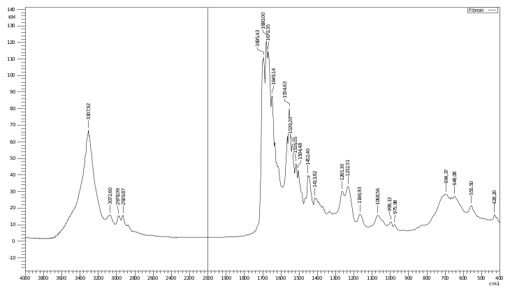
In the spectrum of the initial silk fibroin (Figure 5), the broad intense band around 3319 cm-¹ corresponds to N–H and O–H stretching vibrations belonging to the amide A group. The peaks in the region of 1707–1685 cm-¹ belong to the amide I zone [17,18] and represent the C=O stretching vibrations of the peptide bonds of fibroin. The band observed at 1558 cm-¹ is associated with N–H deformation and C–N stretching vibrations belonging to the amide II group. In addition, signals in the range of 1448–1235 cm-¹ correspond to the vibrations of amide III and C–N and C–O bonds [19,20,21]. The peaks around 1068, 998, 975, 702, and 559 cm-¹ are characteristic of the structural fragments of the fibroin molecule.
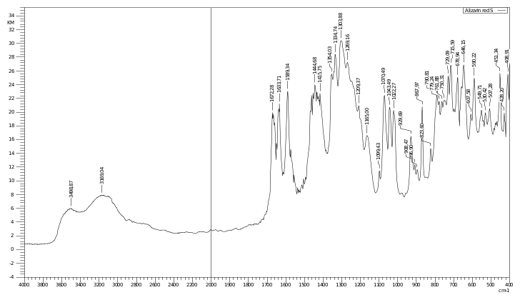
In the FTIR spectrum of Alizarin Red S (Figure 6), broad stretching vibrations of phenolic O–H groups are observed in the regions of 3499 and 3166 cm-¹. The intense peaks in the range of 1672–1639 cm-¹ belong to the carbonyl (C=O) groups of the anthraquinone core. The peaks in the range of 1504–1335 cm-¹ represent C=C vibrations of the aromatic rings and deformation vibrations of the phenolic groups. The intense signals observed in the regions of 1269, 1209, 1165, and 1099 cm-¹ correspond to S=O stretching vibrations of the sulfonate (SO3-) group. Numerous peaks in the low-frequency range of 930–450 cm-¹ reflect out-of-plane deformation vibrations of the aromatic rings [21,22].
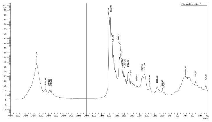
The FTIR spectrum of the silk fibroin sample immobilized with Alizarin Red S (Figure 7) exhibited significant changes compared to the spectrum of the initial fibroin. In particular, the broad band corresponding to O–H/N–H stretching vibrations shifted from 3319 cm-¹ to 3312 cm-¹, which indicates the formation of hydrogen bonds between the hydroxyl groups of the Alizarin Red S molecule and the amine and carbonyl groups of fibroin. The shift of the amide I zone peak from 1707–1685 cm-¹ to the range of 1665–1660 cm-¹ and the change in its intensity indicate an alteration of the microenvironment of the peptide bonds of fibroin. Furthermore, new or intensity-altered signals around 1516, 1452, 1335, 1265, 1225, 1166, and 1068 cm-¹ belong to the aromatic and sulfonate groups of Alizarin Red S, confirming the presence of the reagent on the fibroin surface.
At the same time, the preservation of the main amide I and amide II peaks of fibroin indicates that the primary chemical structure of the fibroin protein was not disrupted during the immobilization of the reagent. The absence of additional intense peaks characteristic of new covalent bonds in the spectra indicates that the immobilization occurred primarily through hydrogen bonds, electrostatic interactions, and π–π intermolecular interactions.
Overall, the FTIR analysis is consistent with the results of diffuse reflectance spectroscopy, confirming the successful immobilization of Alizarin Red S onto the silk fibroin matrix. The preservation of the functional groups of the resulting material indicates the possibility of using it as a sensitive optical sensor material for the sorption-spectroscopic determination of heavy metal ions.
4. Conclusion
In this study, the immobilization of Alizarin Red S onto silk fibroin fiber obtained from local silk waste was investigated, and the optimal conditions of the process were determined. The highest immobilization efficiency was observed at pH 3.32–4.65, a temperature of 25–30 °C, and a duration of 30 min, with an immobilization degree of 78.8 % and a reagent binding capacity of 79.9 mmol·g-¹. UV–Vis, DRS, and FTIR analyses confirmed the successful immobilization of Alizarin Red S onto the fibroin matrix and the preservation of the optical properties of the reagent. The obtained results demonstrate the potential application of the Alizarin Red S–silk fibroin system as a promising optical sensor material for the sorption-spectroscopic determination of heavy metal ions.
References