
Antagonistic activity of endophytic bacteria isolated from Ferula varia against Alternaria SPP
УДК 579
Abstract
For the first time, the taxonomic makeup and functional properties of the endophytic community inhabiting Ferula varia (Schrenk) Trautv. were examined. The work set out to recover endophytes from this plant, describe their morphology, and screen them for the ability to suppress fungal pathogens. Applying a surface-sterilization protocol to the roots, stems, leaves, and flowers of the plant yielded over forty distinct bacterial and fungal endophytes. When these isolates were examined morphologically and stained by the Gram method, most of the bacterial cultures displayed traits typical of the genus Bacillus. Their capacity to restrain fungal growth was screened against the phytopathogen Alternaria spp. by means of the dual-culture technique. Antagonism was recorded in six cultures (FV1i, FV3i, FV8i, FVB1, FVP2 and FVP1.1); of these, FV1i proved the most effective, curbing radial mycelial expansion by as much as 57 %. For this leading strain, the most favourable growth regime was found to combine oat broth as the nutrient base, incubation at 30 °C, and a medium reaction of pH 6.5. Taken together, the data indicate that the endophytes harboured by F. varia and the Bacillus representatives in particular hold appreciable promise as biological agents for managing plant diseases driven by Alternaria.
Аннотация
Состав и биологические свойства эндофитного микробного сообщества, обитающего в Ferula varia (Schrenk) Trautv., были охарактеризованы впервые. Работа была направлена на извлечение эндофитов из данного растения, описание их морфологических признаков и проверку их способности подавлять грибные патогены. При обработке корней, стеблей, листьев и цветков методом поверхностной стерилизации удалось получить свыше сорока различных бактериальных и грибных изолятов. Морфологическое изучение и окрашивание по Граму показали, что большинство бактериальных культур обладают признаками, характерными для рода Bacillus. Их фунгистатический потенциал оценивали в отношении фитопатогена Alternaria spp. с применением метода встречных (двойных) культур. Антагонизм был зафиксирован у шести культур (FV1i, FV3i, FV8i, FVB1, FVP2 и FVP1.1); среди них наиболее результативным оказался штамм FV1i, сдерживавший радиальный рост мицелия вплоть до 57 %. Для этого ведущего штамма оптимальный режим культивирования сочетал овсяный бульон в роли питательной основы, инкубацию при 30 °C и реакцию среды pH 6,5. В совокупности полученные данные говорят о том, что эндофиты F. Varia и в особенности представители рода Bacillus обладают заметным потенциалом в качестве биологических средств борьбы с болезнями растений, вызываемыми Alternaria.
Keywords: F. varia (Schrenk) Trautv; endophytic bacteria; Bacillus; secondary metabolites; antifungal activity; Alternaria spp; dual-culture assay; radial growth inhibition; biocontrol.
Ключевые слова: F. varia (Schrenk) Trautv; эндофитные бактерии; Bacillus; вторичные метаболиты; антифунгальная активность; Alternaria spp; метод двойных культур; ингибирование радиального роста; биологический контроль.
Introduction
Endophytes are microbial partners both bacterial and fungal that reside within plant tissues while leaving the host free of any outward disease signs. Over evolutionary time these microbes have forged a mutually beneficial association with the plants that shelter them, and in doing so they acquire the machinery to manufacture a broad palette of secondary metabolites endowed with antimicrobial, antioxidant, and growth-stimulating activities. The microbial partners of medicinal plants have, over the past several years, drawn growing interest as a fertile reservoir of previously undescribed bioactive molecules and candidate agents for biological control [1-3].
F. varia (Schrenk) Trautv. is a plant of regional character, occurring in the wild across Uzbekistan, Kazakhstan, and Kyrgyzstan. Members of the genus Ferula have a long record of folk-medical use as remedies that relieve spasms, expel gas, and act as antiseptics largely thanks to the abundance of bioactive terpenoids and coumarins they accumulate. Despite this, the microbial endophytes of the genus have received little scientific attention. Illustratively, a survey of more than 170 bacterial endophytes extracted from Ferula songorica showed that most of them fell within the phylum Actinobacteria [4], As far as we are aware, no findings on the endophytic microflora of F. varia (Schrenk) Trautv. have yet appeared in the literature.
The genus Alternaria, meanwhile, ranks among the most damaging groups of plant-pathogenic fungi across the globe. Its members provoke an assortment of disorders leaf spotting and fruit decay among them in upwards of 400 host species, translating into heavy economic damage in the agricultural sector [5]. Synthetic fungicides continue to serve as the mainstay of control, yet their harmful ecological footprint, together with the steady rise of fungicide-tolerant strains, has made the search for greener control options a pressing priority. Against this backdrop, bacterial endophytes especially those belonging to the genus Bacillus stand out as attractive biocontrol candidates, chiefly because they secrete antifungal lipopeptides of the surfactin, iturin, and fengycin families.
Given these considerations, the present investigation was designed to recover endophytic microorganisms from F. varia (Schrenk) Trautv., to profile them according to their morphological traits, and to gauge their fungistatic performance against the phytopathogen Alternaria spp. through the dual-culture approach. A further objective was to establish, for the most active isolate, the culturing regime nutrient medium, incubation temperature, and pH that would best drive the output of its bioactive metabolites.
Materials and Methods
Plant Material
Sampling was carried out in the first days of May 2025 in the Konimex district of the Navoiy Region, Uzbekistan (40.739287, 63.747228). Vigorous, disease-free F. varia (Schrenk) Trautv. specimens were dug up with care so as not to damage the root architecture and were brought back to the laboratory along with the adhering rhizosphere soil [6].
Isolation of Endophytic Microorganisms
The stems, foliage, roots, and floral parts of F. varia (Schrenk) Trautv. were passed through a surface-disinfection sequence. To begin with, the tissues were rinsed at length beneath running tap water and then flushed with sterile distilled water. They were next held in 70 % ethanol for 2 min, transferred to a 10 % sodium hypochlorite bath for a further 2 min, and finally washed five successive times in sterile distilled water. As a check on the disinfection step, a 100 µL portion of the water from the fifth rinse was spread over Nutrient Agar (NA) and incubated to confirm that no colonies developed.
Once surface-sterilized, the samples were left to dry on sterile paper towels for 5 min under aseptic conditions. Roughly 10 g of the fresh, sterile tissue was then ground in a sterile porcelain mortar. A 1 g portion of the resulting homogenate was suspended in 9 mL of sterile distilled water and blended thoroughly on a vortex mixer, after which the suspension was subjected to a serial dilution down to 10⁻⁵.
Measured volumes of the relevant dilutions were spread onto Nutrient Agar (NA; peptone, 5 g L⁻¹; yeast extract, 3 g L⁻¹; NaCl, 5 g L⁻¹; agar, 15 g L⁻¹; pH 7.4) to recover bacteria, and onto Potato Dextrose Agar (PDA; potato infusion from 200 g potatoes L⁻¹; dextrose, 20 g L⁻¹; agar, 15 g L⁻¹; with streptomycin added at 50 mg L⁻¹ after autoclaving; pH 5.7) to recover fungi. Plates were then held at 28 °C for 5 days.
Colonies that differed in shape, pigmentation, or texture were picked individually and repeatedly re-streaked on fresh NA or PDA until pure cultures were obtained. The purified isolates underwent an additional 5 day incubation at 28 °C prior to the analyses that followed [7-10].
Antifungal Activity Assay
The ability of the bacterial endophytes to antagonize phytopathogenic fungi was tested in vitro by the dual-culture method against Alternaria spp. Discs of agar 6 mm across were cut aseptically from the advancing edge of a 7 day old Alternaria spp. colony with a sterile cork borer and set at the middle of freshly poured Potato Dextrose Agar (PDA) plates.
In the treatment plates, 18-24 h cultures of the bacterial endophytes were introduced onto the same PDA surface by the agar-plug technique at two diametrically opposite points, some 35-40 mm away from the fungal disc. Control plates carried only the Alternaria spp. disc at the centre, with no bacterial inoculum. Every treatment was set up in triplicate.
Incubation proceeded in darkness at 25 – 28 °C for 7 – 10 days, continuing until the fungal colony on the control plates had spread over nearly the whole Petri dish. Afterwards, the radial extension of the fungal colony was recorded in millimetres along two axes toward the bacterial streak and directly away from it with a sterile ruler [11, 12].
To place the antagonism on a quantitative footing, the radial-inhibition percentage (RI, %) was derived from the expression below:
in which R1 denotes the radius of the pathogen colony on the control plate (grown without any antagonist), and R2 the radius of the pathogen colony on the treatment plate (grown in the company of the endophyte under test). Using the RI (%) figures obtained, each isolate was assigned to an antagonism category according to the thresholds set out in Table 1. Results are reported as mean ± standard deviation (M ± SD) from three independent replicates. Differences between groups were judged by one-way analysis of variance (ANOVA) with Tukey's post-hoc comparison, treating p < 0.05 as significant [13, 14].
Table 1. Categories used to grade the strength of antagonism from radial-inhibition values (RI, %).
| Radial inhibition (RI, %) | Antagonistic Activity |
|---|---|
| ≥ 75 | High (strong) antagonism |
| 50–74 | Moderate antagonism |
| 25–49 | Weak antagonism |
| < 25 | No detectable antagonism |
Optimization of Cultivation Conditions
For strain FV1i, which had shown the strongest antifungal response, the growth conditions were fine-tuned by testing how the choice of medium, the incubation temperature, and the pH influenced both biomass accumulation and the yield of bioactive metabolites. The strain was propagated in a panel of liquid media Nutrient Broth (NB), Nitrogen Free Broth (N-free broth), Oat Broth, Luria Bertani (LB) Broth, and Tryptic Soy Dextrose (TSD) Broth across a range of temperatures and pH values (Table 2). Growth and metabolite output were monitored under each setting, and the medium that gave FV1i the greatest biomass together with the highest metabolite production was adopted as optimal for all later work [1, 7, 15].
Table 2. Composition of the culture media used for optimization of the cultivation conditions of strain FV1i.
| Medium | Composition (g/L) | pH |
|---|---|---|
| Nutrient broth (NB) | Peptone, 5.0; beef extract, 3.0; NaCl, 5.0 | 7.4 ± 0.2 |
| Nitrogen-free broth (N-free broth) | Mannitol, 20.0; K₂HPO₄, 0.5; MgSO₄·7H₂O, 0.2; NaCl, 0.2; CaSO₄, 0.1; CaCO₃, 5.0; trace elements | 7.0 ± 0.2 |
| Oat broth | Oat infusion (from 50 g oat flakes); glucose, 20.0 | 6.5 ± 0.2 |
| Luria–Bertani (LB) broth | Tryptone, 10.0; yeast extract, 5.0; NaCl, 10.0 | 7.0 ± 0.2 |
| Tryptic soy dextrose (TSD) broth | Pancreatic digest of casein, 17.0; papaic digest of soybean meal, 3.0; NaCl, 5.0; K₂HPO₄, 2.5; dextrose, 2.5 | 7.3 ± 0.2 |
Preparation of Bacterial Extracts
Following incubation, the bacterial culture was spun at 4,000 rpm for 20 min and the cell-free supernatant recovered. This supernatant was partitioned against ethyl acetate at a 1:1 ratio in a separating funnel. Once the phases had separated, the organic layer was drawn off and taken to dryness under reduced pressure on a rotary evaporator, yielding the crude bacterial extract [16-18].
Statistical Analysis
Following incubation, the bacterial culture was spun at 4,000 rpm for 20 min and the cell-free supernatant recovered. This supernatant was partitioned against ethyl acetate at a 1:1 ratio in a separating funnel. Once the phases had separated, the organic layer was drawn off and taken to dryness under reduced pressure on a rotary evaporator, yielding the crude bacterial extract [19, 20].
Results and discussion
Isolation and Morphological Characterization of Endophytic Microorganisms
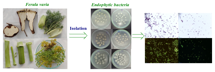
In all, more than forty bacterial and fungal endophytes were retrieved from the roots, stems, leaves, and flowers of F. varia (Schrenk) Trautv. As a rule the bacterial colonies were sizeable, with ragged edges and surfaces that were either coarse or waxy. The prevailing colours ranged from white to cream, though a handful of isolates gave off characteristic pigments. Colony textures spanned dry to moist, and a number of cultures took on a rhizoid outline. Gram staining classified every bacterial isolate as Gram-positive. On the combined evidence of colony appearance and Gram reaction, the bacterial isolates were provisionally referred to the genus Bacillus. That Bacillus spp. should dominate this endophytic assemblage fits well with the genus's recognized knack for producing endospores and withstanding environmental stress attributes that let it settle and endure inside plant tissues more successfully than many competing bacterial lineages.
Antifungal Activity
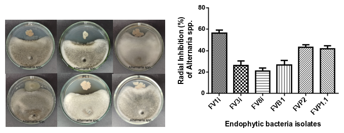
The fungistatic performance of the bacterial endophytes toward Alternaria spp. was assessed by the dual-culture assay. Out of the whole panel, only six isolates displayed antagonism against the pathogen. Pride of place went to isolate FV1i, which held back the radial spread of Alternaria spp. by 57 % a level that qualifies as strong antagonism. Isolates FVP2 and FVP1.1 likewise proved noteworthy, each trimming radial growth by 43 %, a figure that places them in the moderate-antagonism bracket. By contrast, FV3i and FVB1 curtailed fungal growth by 28 %, and FV8i by 21 %, so these three were rated as weak antagonists. On balance, FV1i emerged as the isolate with the greatest antagonistic capacity against Alternaria spp. and was therefore carried forward into the optimization phase aimed at boosting bioactive-metabolite production.
Optimization of the Most Active Strain
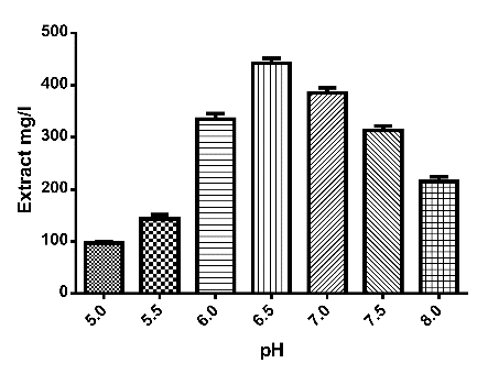
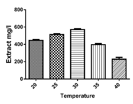
In light of the antifungal screening, isolate FV1i stood out as the top performer and was accordingly chosen for optimization, in which the impact of assorted media, incubation temperatures, and pH levels on growth and metabolite formation was explored. Of all the regimes trialled, FV1i turned out the largest quantity of bioactive metabolites when cultured in oat broth at 30 °C and pH 6.5. These settings were consequently taken as optimal for growing strain FV1i and were retained for the remaining experiments.
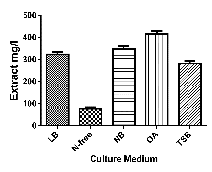
The dominance of Bacillus within the endophytic community of F. varia (Schrenk) Trautv. echoes earlier accounts of endophytic populations described from other plant hosts. This genus is widely noted both for its ability to establish stable residence in plant tissues and for the varied assortment of bioactive secondary metabolites it generates. Because the antifungal capacity of endophytes tied to F. varia has not previously been examined in any depth, the 57 % radial suppression of Alternaria spp. achieved by strain FV1i stands out as a meaningful result. Such antagonism is most plausibly linked to the secretion of antifungal lipopeptides surfactin, iturin, and fengycin among them which are known to compromise the integrity of the fungal cell membrane and, in turn, to arrest fungal development.
The optimal culturing conditions pinpointed for strain FV1i oat broth, an incubation temperature of 30 °C, and pH 6.5 align with the settings reported to favour secondary-metabolite formation in Bacillus species; such conditions have been tied to elevated expression of the genes underpinning antimicrobial compound biosynthesis. On the whole, the present results point to strain FV1i as a strong candidate for the biological management of diseases attributable to Alternaria spp. Even so, additional work will be needed to isolate and characterize the precise bioactive constituents behind its antifungal effect and to unravel the mechanisms through which they act.
Conclusion
The present study yielded more than forty endophytic microbial isolates from the roots, stems, leaves, and flowers of F. varia (Schrenk) Trautv. On morphological grounds, the bulk of the bacterial isolates were tentatively assigned to the genus Bacillus. Dual-culture screening identified six isolates with antifungal action against Alternaria spp., strain FV1i being the most inhibitory at 57 %. The culturing regime best suited to FV1i was established as oat broth, incubation at 30 °C, and pH 6.5.
Collectively, these observations underscore the promise of the F. varia endophytic microflora strain FV1i above all as a candidate biological agent for countering Alternaria-associated fungal diseases. The study likewise lays the groundwork for subsequent efforts directed at pinpointing the bioactive metabolites this strain produces and at assessing how they might be applied within sustainable plant-disease management.
References

Doctor of Biological Sciences, Professor,
Chief Research Officer Institute of Microbiology, Academy of Sciences of the Republic of Uzbekistan,
Uzbekistan, Tashkent
E-mail: k-davranov@mail.ru